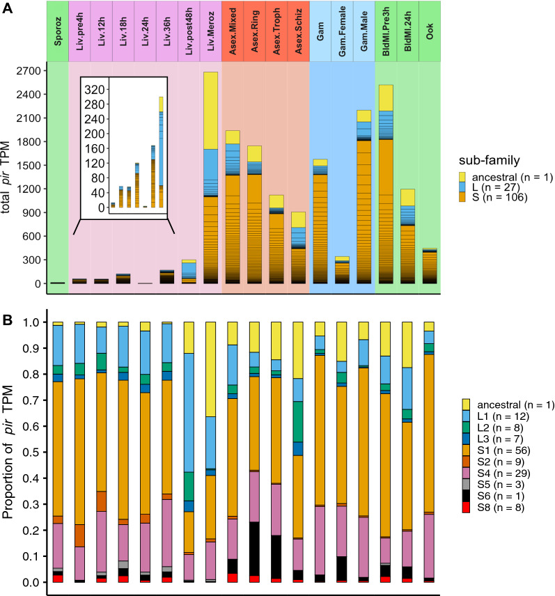
Fig. 2.
Expression of pir genes throughout P. berghei life cycle. A Bar chart of total pir gene TPM (y-axis), calculated from the mean expression across all experimental samples of each life cycle stage (x-axis). The colours of the bar chart denote the classification; ancestral (yellow), Long (L) (blue) and Short (S) (orange), with the boxes of the stacks denoting expression of the individual genes. The background and strip name colours correspond to groups of life cycle stages, including mosquito (green), liver (pink), asexual blood (red), and gametocyte (blue) stages. The genes in each subfamily are ordered by expression levels. Inset of figure shows an enlargement of the sporozoite and liver stages. B Bar chart of the proportion of total pir gene TPM (y-axis), contributed by the ancestral pir (yellow) and family sub-divisions of the Short/Long pirs, for each stage (x-axis). Stages are separated as in (A). Legend shows the number of genes that are members of each grouping, which are collated together for each box in the stacked bars. The ordering is by family sub-division, from ancestral, to Long groups (1–3) and finally Short groups (1–2, 4–6, 8)

