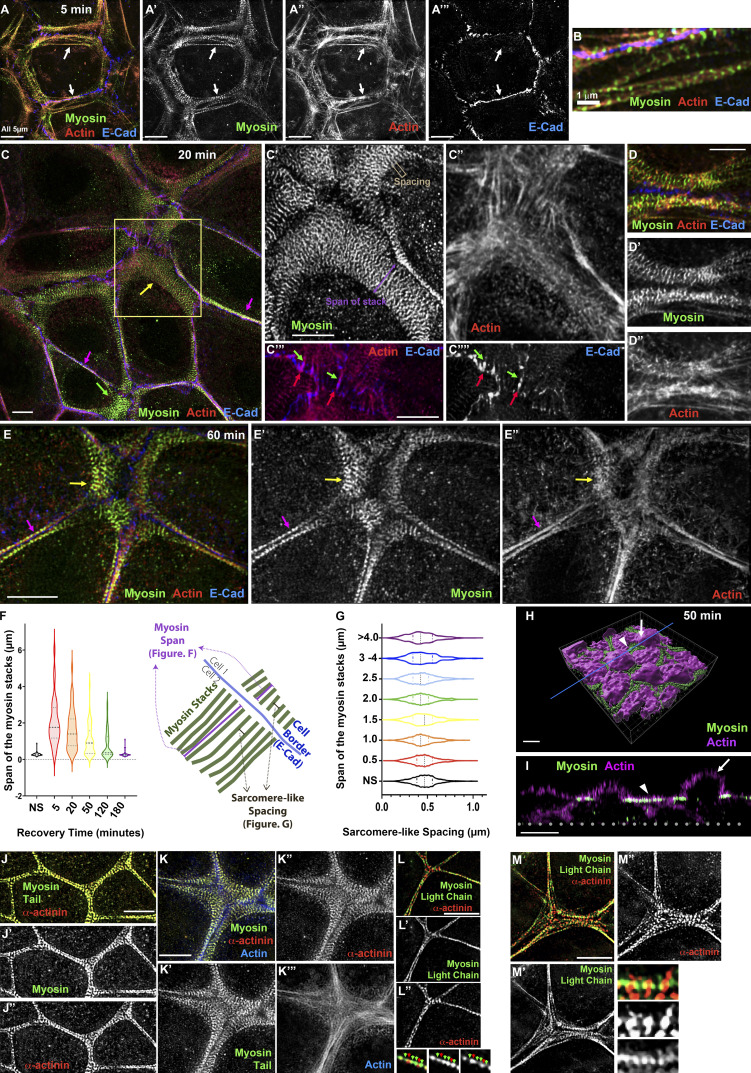
Figure 2.
Extensive stacks of myosin precede the formation of bundled actin as Ecad–based AJs assemble. (A–E) Images collected at different time points of Ca recovery reveal that an extensive myosin array forms during midrecovery and localizes with a less well-organized actin network. (F and G) Quantification. Despite changes in the span of the myosin stacks at different time points (F), spacing between myosin stacks remains similar (G). (H) 3D surface rendering of a cell in midrecovery. (I) Cross-section view at the line indicated in H. The extensive myosin stacks localize to a restricted Z plane underlying the apical plasma membrane (arrowheads), between the domed microvillar caps (arrows). In F, n = (nonswitched [NS] = 75, 5 min = 34, 20 min = 44, 50 min = 63, 120 min = 51, 180 min = 43). In G, n = 550 for each time point.

