Figure S4.
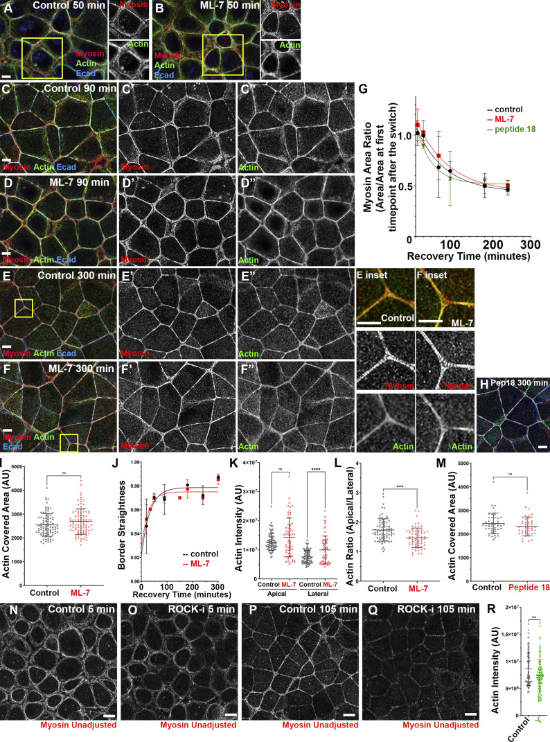
Inhibiting MLCK using ML-7 or peptide-18 does not alter myosin localization during recovery or prevent or delay assembly of the final ZA actomyosin structure. (A–F) Representative images showing cells in control vs. ML-7–treated cells at different time points during Ca recovery. By the last time point, cells assembled bundled F-actin decorated by sarcomeric myosin in both control and drug-treated conditions (E vs. F; H). (G and I–M) Quantification. Myosin maturation (G), actin bundling at the ZA (I and M), and border straightening (J) were similar in control and drug-treated conditions. After ML-7 treatment, there was some elevation of lateral actin and thus apical actin polarization was reduced (L). (N–Q) Representative images showing the difference in apical myosin signal in control (N and P) vs. ROCK-inhibited (O and Q) conditions. Myosin signal at the junction is reduced when ROCK is inhibited. (R) Quantification. Levels of apical actin were reduced after ROCK inhibition. In G, numbers for each time point are in Table S1. In I, K–M, and R, n = individual borders; control and ML7 = 94 (I); control = 80, ML7 = 63 (K and L); control = 45, peptide 18 = 43 (M); and control = 95, Y = 85 (R). In J, representative of five experiments with two fields of cells/experiment/time point, with seven to nine borders quantified/field. Statistical analysis was performed with unpaired two-way t tests (M) or one-way ANOVA tests and post hoc Tukey tests (I, K, L, and R). Error bars represent mean ± SD. ****, P < 0.0001; ***, P < 0.001; **, P < 0.01. Boxes indicate areas magnified at right.

