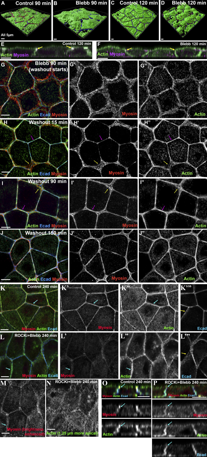
Figure S5.
3D cell shapes after blebbistatin treatment, blebbistatin washout restores the ZA, and dual inhibition of both ROCK and the myosin ATPase. (A–D) 3D images of cell surface in control vs. blebbistatin-treated cells. The apical surface of the control cells becomes flatter as junctions mature (A vs. C; also in Fig. S1, M–Q). In contrast, blebbistatin-treated cells were not able to flatten the apical surface, even at the later recovery time points (B and D). (E and F) Cross-section views. After blebbistatin treatment, cells still retained the ability to regain a more columnar architecture as lateral borders zipped up, but actin and myosin were not apically polarized (E vs. F, arrows). (G–J) Representative images showing the recovery of ZA actomyosin structures after blebbistatin washout. Within 15 min after washout, actin and myosin already began to focus at bicellular borders (H′ and H″, magenta arrows). Myosin at the tricellular borders was slower to recover (H and I, yellow arrows), but by 150 min (J), ZA actomyosin structures returned to those seen in the control mature monolayer. (K–P) Combined inhibition of ROCK and blebbistatin does not prevent Ecad-based adhesion but does disrupt ZA assembly and Ecad polarization. (K and L) Combined treatment reduces junctional myosin, actin bundling at the ZA, and Ecad apical polarization (K″′ vs. L″′). (M) Dual treatment with myosin signal enhanced. (N) After dual treatment, spiky actin covers the apical surface. (O and P) Cross sections. Ecad-based junctions zip up, but apical enrichment of actin and Ecad is reduced (arrows).

