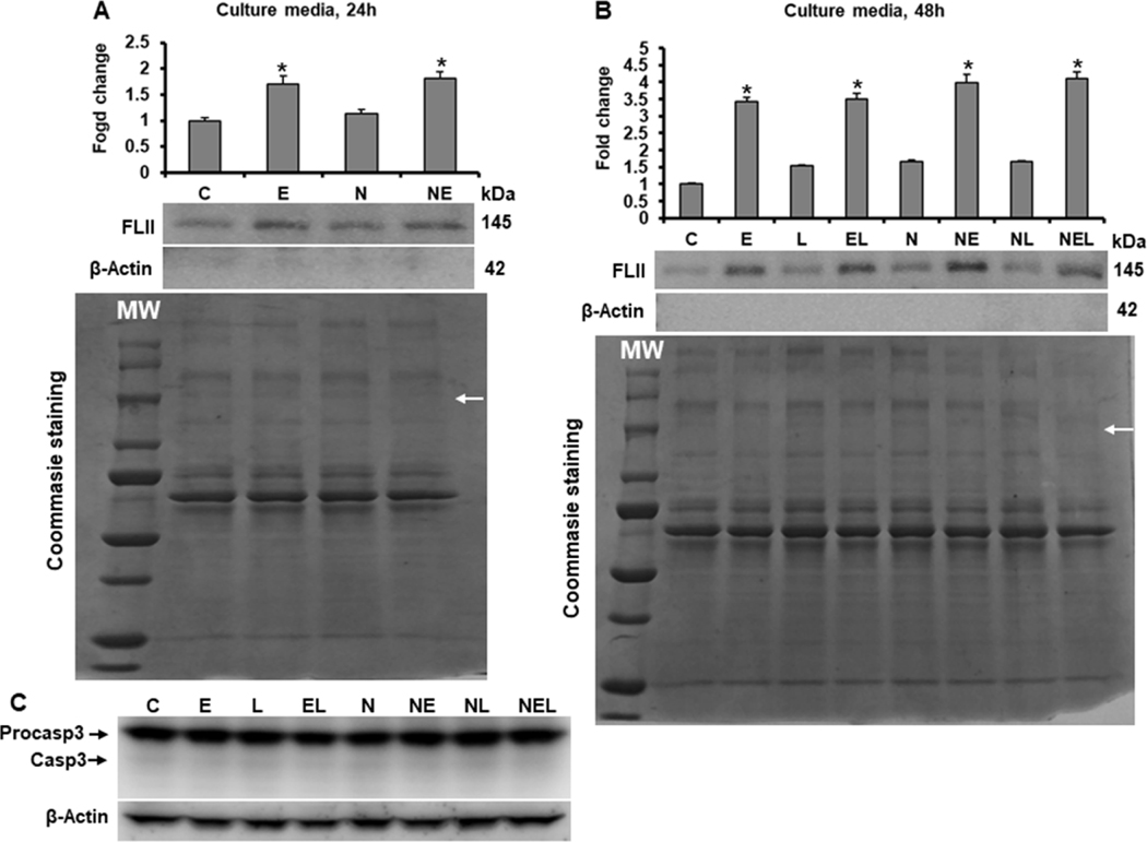
Fig. 7. Effect of ethanol on FLII secretion into cell culture medium.
Determination of FLII secretion after (A) 24 and (B) 48 h of ethanol and/or LPS exposition. Culture media were collected at indicated times and prepared as described in materials and methods section. β-Actin was detected to discard that secretion of FLII was due to cells breaking. Protein levels were normalized to Coommasie Blue-stained gels used as loading control. (C) Cleaved Caspase-3 (Casp3) was detected in total protein from co-cultures to discard that secretion of FLII was due to programmed cell death activation. Bars values are expressed as fold change compared to controls and represent the mean ±SE. n=6 group. Statistically different from *C group; p<0.05. White MW letters and arrows on Coommasie Blue-stained gels indicate protein Molecular Weights and FLII size location, respectively. Groups: C, Control; E, Ethanol; L, LPS; and EL, Ethanol plus LPS, were transfected with EV and were called untransfected cells. N, NXN-transfected cells; these cells were also exposed to E, L and EL treatments and were identified as: NE, NL and NEL, respectively.

