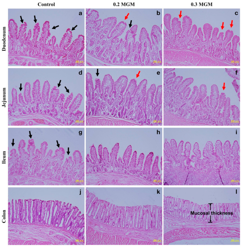
Figure 6.
Representative histological micrographs of the duodenum, jejunum, ileum, and colon weaned piglets at 20 days post-weaning, produced by hematoxylin and eosin staining. Scale bar, 200 μm. Abbreviations: Control, control group; 0.2 MGM, 0.2% MGM-P group; 0.3 MGM, 0.3% MGM-P group. Pathological changes at the tips of intestinal villi during weaning are indicated by red (slight damage) and black (serious damage) arrows. The exposed lamina propria are clearly visible in the control group (a,d,g). The mucosal integrity is relatively high in the treatment group (b,c,e,f,h,i) especially in 0.3 MGM group (f,h,i). The mucosal thickness of the colon is relatively thick in the control group (j) and 0.2 MGM group (k) than that in the 0.3 MGM group (l) (including the muscularis mucosae).

