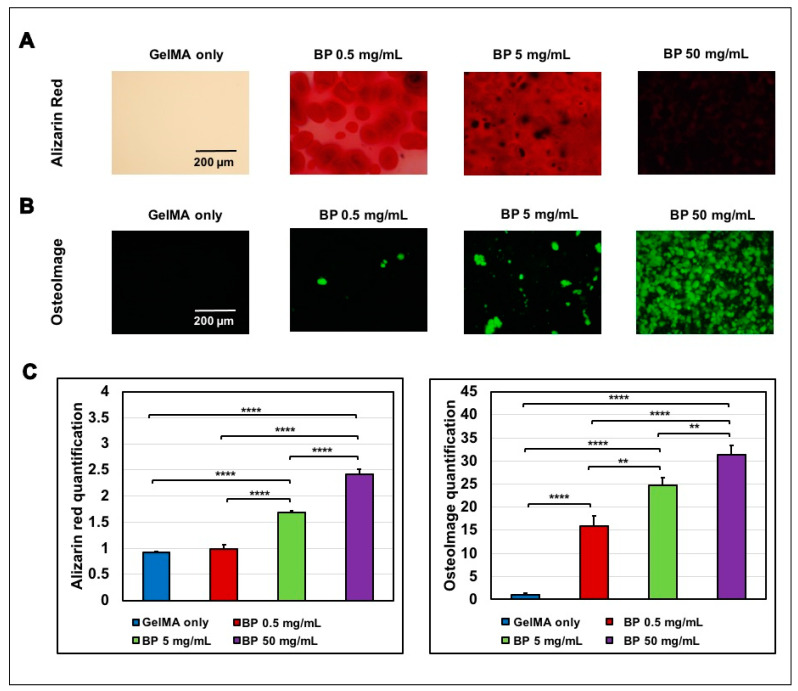
Figure 2.
Evaluation of the calcium and hydroxyapatite components in the GelMA/BP composite hydrogels. The alizarin red images were taken by staining scaffolds without cells so that the alizarin red dye stains calcium content due to the presence of BP within the scaffolds. (A) Alizarin Red staining of calcium-rich portions (Scale bar: 200µm). (B) Osteoimage staining of hydroxyapatite portion in green (Scale bar: 200µm). (C) Quantification of the red color intensity in Alizarin Red staining (Error bars: ±SD, ** p < 0.01, **** p < 0.0001) and quantification of the green fluorescence in Osteoimage staining.

