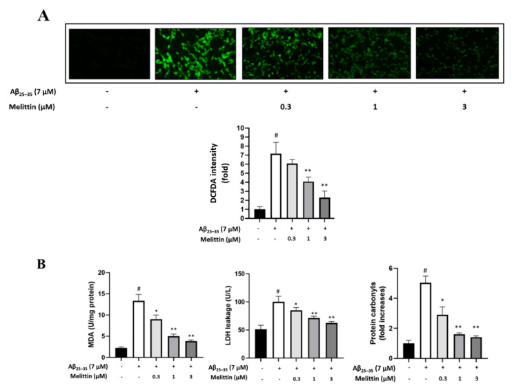
Figure 3.
Melittin regulates cellular oxidative stress induced by Aβ25–35 in HT22 cells. (A) Immunofluorescence analysis to determine the cellular ROS rate. The result indicated that melittin at 0.3 to 3 μM dosage-dependently normalized the expression of pro and anti-apoptosis proteins under Aβ25–35 stress challenge. To conduct this experiment: Seven hours after Aβ25–35 (7 μM) challenge in 96-well plates, immunofluorescence analysis by DCFDA staining was carried out. (B) Cellular MDA, LDH, and protein carbonyls levels. The result indicated that melittin at 0.3 to 3 μM dosage-dependently down-regulated MDA, LDH, and protein carbonyl parameters under Aβ25–35 stress challenge. To conduct this experiment: Seven hours after Aβ25–35 (7 μM) challenge in 6-well plates, kits measuring MDA, LDH, and protein carbonyls were used to determine the parameters. Data are presented as mean ± standard deviation values of triple determinations. # p < 0.01 vs. control * p < 0.05 and ** p < 0.01 vs. Aβ25–35 only-treated group.

