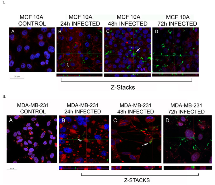
Figure 1.
Representative confocal microscopy images of MCF 10A (Panel I) and MDA-MB-231 (Panel II) mammalian epithelial cells infected with B. burgdorferi. The cells were co-cultured with B. burgdorferi for (B) 24 h, (C) 48 h, and (D) 72 h. (A) Uninfected cells treated only with co-culture media were used as a control. CM-Dil membrane stain was used to label the cells (red), DAPI was used to stain the nucleus (blue) and polyclonal anti-B. burgdorferi was used to visualize B. burgdorferi cells (green), as described in the Material and Methods. The representative images were taken at 630X with a scale bar of 20 μm. White arrowhead represents an invading spirochete and white arrow shows small B. burgdorferi aggregates inside the cells.

