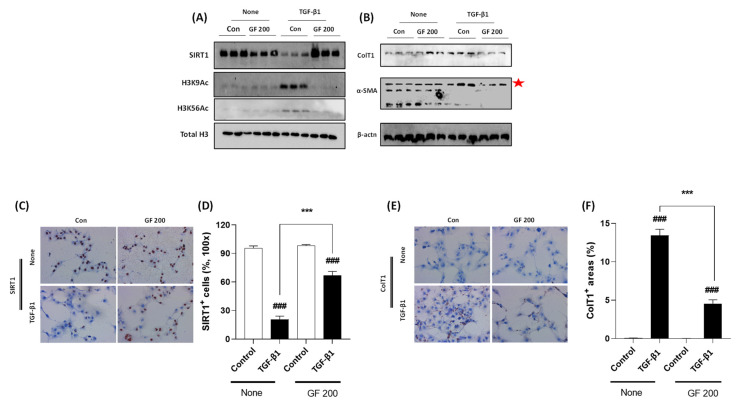
Figure 7.
GF Alleviates TGF-β1-induced LX-2 Cell Activation via Enhancement of SIRT1. Western blot analysis in TGF-β1 treated LX-2 cells with or without GF. (A) Cellular protein levels of SIRT1, H3K9Ac, H3K56Ac, and Total H3. (B) Cellular protein levels of Collagen type 1, α-SMA, and β-actin. IHC analysis against SIRT1 (C) and its quantification analysis (D), and Collagen type 1 IHC (E) and its quantification analysis (F). Data were expressed mean ± SEM (n = 3 for Western blot analysis, n = 4 for IHC analysis). ### p < 0.001 for Control vs. TGF-β1 and *** p < 0.001 for None vs. GF 200. IHC images were captured by microscope condition (100× magnifications).

