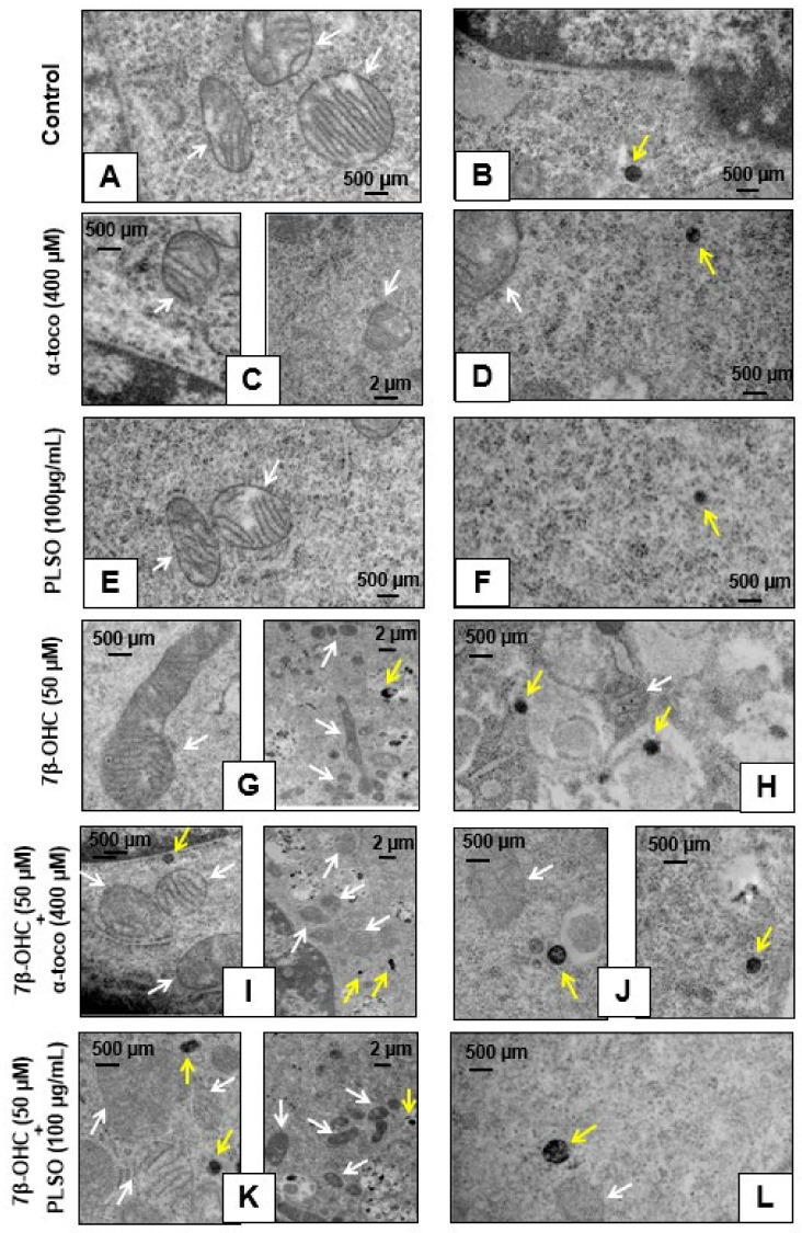
Figure 10.
Visualization of the mitochondria and peroxisomes in C2C12 myoblasts by transmission electron microscopy. C2C12 cells were incubated for 24 h with or without 7β-OHC (50 µM) in the presence or absence of PLSO (100 µg/mL) or α-tocopherol (400 µM). In untreated cells (control) (A,B), α-tocopherol (400 mM)-treated cells (C,D), and PLSO (100 µg/mL)-treated cells (E,F), numerous mitochondria with clear cristae as well as round and regular peroxisomes were detected. In 7β-OHC (50 µM)-treated cells (G,H), irregular mitochondria with an increased size, reduced matrix density, and disrupted cristae, as well as peroxisomes with abnormal sizes and shapes were visualized. In (7β-OHC + α-tocopherol)-treated (I,J) and (7β-OHC+ PLSO) (K,L)-treated cells, mainly mitochondria and peroxisomes morphologically similar than those present in the control cells were observed. The white arrows point towards mitochondria and the yellow arrows point towards peroxisomes.

