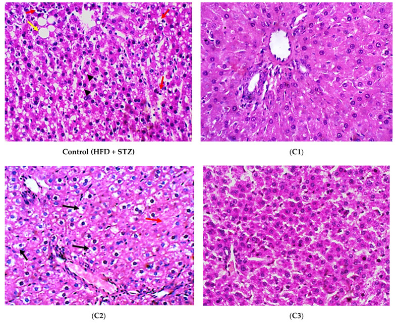
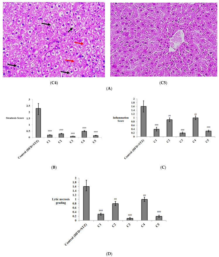
Figure 5.
The histopathological study of hepatic tissues from different study groups. (A) Photomicrographs of hepatic tissues from different experimental groups stained by hematoxylin and eosin (H&E) and examined with magnifying power (40×). HFD + STZ control group showed hepatocytes with micro-vesicular steatosis (arrow heads), as well as macro-vesicular steatosis (yellow arrow). There were scattered foci of lytic necrosis of hepatocytes (red arrows) (H&E, 40×). (C1): Rutin-treated group showed uniform hepatocytes with no evidence of injury (H&E, 40×). (C2): Quercetin 3-O-neohesperdoside-treated group: Hepatocytes showed no evidence of steatosis, yet, several hepatocytes still showed hydropic degeneration (black arrows), with few foci of lytic necrosis (red arrows) (H&E, 40×), (C3): Quercetin-3-O-β-galactoside-treated group, in which hepatocytes showed no evidence of steatosis or hydropic degeneration. No other pathological changes observed (H&E, 40×), (C4): Isoquercetrin-treated group; in which hepatocytes showed no evidence of steatosis, yet, several hepatocytes still show hydropic degeneration (black arrows), with few loci of lytic necrosis (red arrows) (H&E, 40×). (C5): Quercetin-treated group; in which hepatocytes showed no evidence of steatosis or hydropic degeneration. No other pathological changes observed (H&E, 40×), (B–D) histopathological scores of liver steatosis, inflammation, and lytic necrosis, respectively. Data are expressed as mean ± SD and analyzed using one-way ANOVA followed by Bonferroni’s post hoc test. ## significantly different compared to the T2DM control group (high fat diet + Streptozotocin) at at p < 0.01; ### at p < 0.001.


