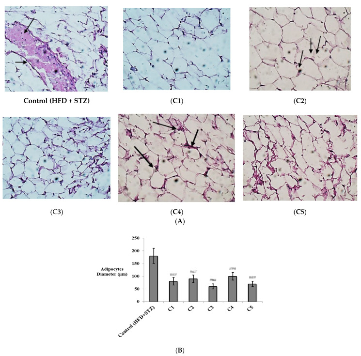
Figure 6.
The histopathological study of adipose tissues from different study groups. (A) Photomicrographs of adipose tissues from different experimental groups stained by hematoxylin and eosin (H&E) and examined with magnifying power (40×). HFD + STZ control group; adipose tissue showed many congested vessels (Black arrows) (H&E, 40×). (C1): Rutin-treated group; adipose tissue showed uniform fat cells, with non-congested blood vessels (H&E, 40×). (C2): Quercetin 3-O-neohesperdoside-treated group; adipose tissue showed few congested vessels (Black arrows). (C3): Quercetin-3-O-β-galactoside-treated group; adipose tissue showed uniform fat cells, with non-congested blood vessels. No inflammatory changes observed (H&E, 40×), (C4): Isoquercetrin-treated group; adipose tissue showed few congested vessels (Black arrows) (H&E, 40×), (C5): Quercetin-treated group; adipose tissue showed uniform fat cells, with non-congested blood vessels (H&E, 40×). No inflammatory changes observed. (B) Diameter of adipocytes (µm) expressed as mean ± SD and analyzed using one-way ANOVA followed by Bonferroni’s post hoc test. ### significantly different compared to at p < 0.001.

