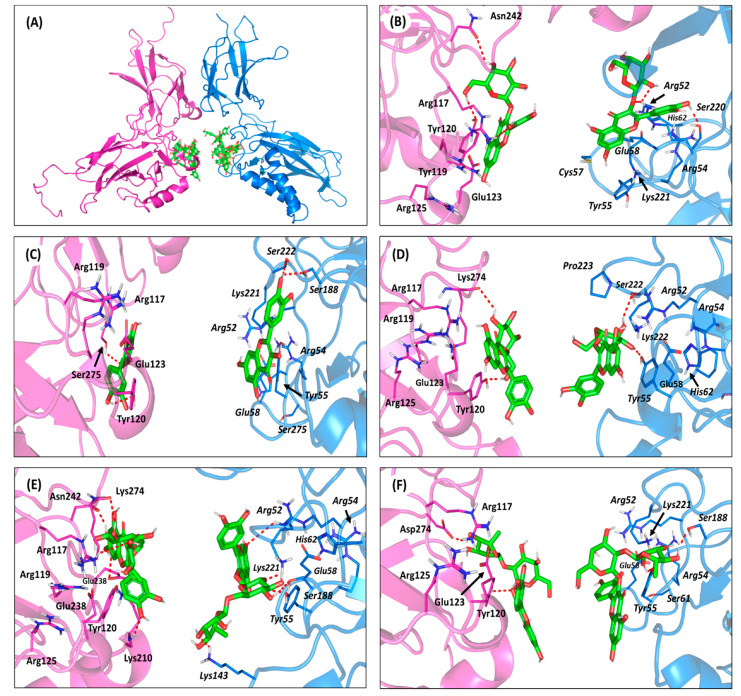
Figure 7.
Molecular docking investigation of the isolated compounds at DNA-binding domains of the NF-κB RelB:p52 heterodimer (PDB ID: 3DO7). (A) An overlay of the docked ligands (green sticks) at both NF-κB heterodimer subunits, RelB (magenta cartoon) and p52 (marine blue cartoon). (B–F) The predicted binding modes of quercetin-3-O-β-galactoside(B); quercetin (C); isoquercitrin(D); rutin(E); and quercetin-3-O-neohesperdoside (F) at RelB (left side) and p52 (right side) illustrated as cartoon 3D-representation within their respective magenta and marine blue colors. Polar interactions, represented as hydrogen bonds, are illustrated as red dashed lines and only residues (lines), located within a 4 Å radius of bound ligand, are displayed and labeled with sequence number.

