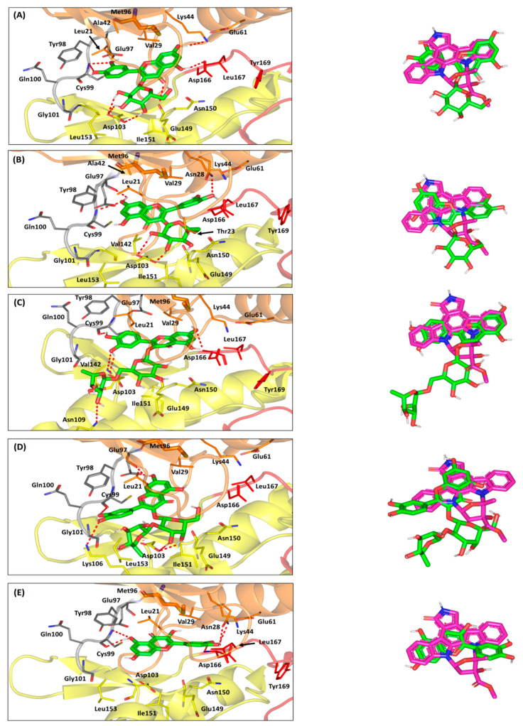
Figure 8.
Molecular docking investigation of the isolated compounds at ATP-binding site of the hIKK protein target (PDB ID: 4KIK). Left panels illustrate the proposed docking poses for the investigated compounds (green sticks) at the catalyric ATP-binding site of IKK target protein (orange, yellow, gray, and red cartoons for N-lobe, C-lobe, hinge region, and activation segment, respectively); (A) quercetin-3-O-β-galactoside; (B) isoquercitrin; (C) rutin; (D) quercetin-3-O-neohesperdoside; (E) quercetin. On the right, overlay of docked isolated compounds (green sticks) and crystallized ligand, KSA (magenta sticks), depicting their comparative orientations within the catalytic binding site. Polar interactions, represented as hydrogen bonds, are illustrated as red dashed-lines and only residues (lines), located within 4 Å radius of bound ligand, are displayed, labeled with sequence number, and colored based on their respective position.

