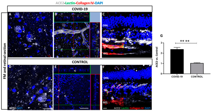
Figure 3.
Angiotensin-converting enzyme 2 (ACE2) expression in COVID-19 (A–C) and control retinal vessels (D–F). ACE2 expression (white) observed in retinal ganglion cells (RGCs) in superficial vasculature in COVID-19 (A) and control (D) retinas. ACE2 is observed in cells in inner nuclear layer (INL) and ganglion cell layer (GCL) and endothelial cells in vessels of COVID-19 (B) and controls (E). ACE2 localization in Optimal compound tissue (OCT)-embedded retinas cross-sections previously labelled with lectin (green) and collagen IV (red) in COVID-19 (C) and controls (F). B and E represent an orthogonal projection of retinal vessels. 4′,6-diamidino-2-phenylindole (DAPI) (blue) label nuclei. (G) Quantification of immunofluorescence intensity in percentage of COVID-19 retinas vs. controls. **** p < 0.0001. Scale bar: 100 µm. Abbreviations: GCL (ganglion cell layer), INL (inner nuclear layer), ONL (outer nuclear layer).

