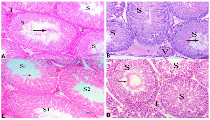
Figure 6.
Histopathological characteristics of seminiferous tubules of testes from the four studied rat groups (A–D) using hematoxylin and eosin (H and E) staining and magnification ×200. (A) Control group appearance with normal seminiferous tubules (S) containing normal spermatogenic cells and spermatid as well as normal interstitial tissues (I) (B) CdCl2 group showing seminiferous tubules (S) lined with degenerated spermatogenic cells and low amounts of spermatid and sperms (arrow). Note, the interstitial tissues contain congested blood vessels (V) and inactive Leydig cells. (C) CdCl2 and Vit. E group show normal seminiferous tubules (S1). The other tubules (S2) are lined with degenerated spermatogenic cells with a few sperms. The interstitial tissues contain normal blood capillaries and less active Leydig cells (I). (D) CdCl2 and RCME group show normal seminiferous tubules (S) with spermatogenic cells and sperms (arrow). The interstitial tissues (I) appear normal.

