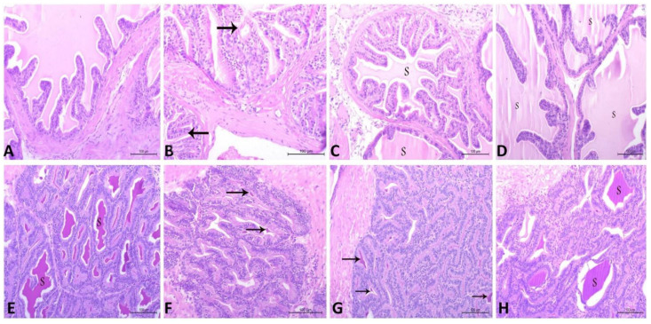
Figure 7.
Histopathological characteristics of prostatic tissue (A–D) and vesicular gland (E–H) in adult male albino rats using hematoxylin and eosin (H and E) staining and magnification ×200. (A) Control group containing normal prostatic acini, lined with simple columnar secretory epithelium with secretory materials. (B) CdCl2 group showing prostatic acini, which appear collapsed and inactive and are separated by thick connective tissue. The acini are lined with low columnar epithelium with low secretory activity. Note, few secretory materials appear in the lumen of the prostatic acini (arrow). (C) CdCl2 and Vit. E group show less active prostatic acini lined with less active secretory cells and few amounts of secretory materials (S). (D) CdCl2 and RCME group show normal prostatic acini lined with simple columnar secretory epithelium, with secretory materials (S). (E) Control group contains normal vesicular gland acini lined with high columnar epithelium, with huge amounts of secretory materials (S). (F) CdCl2 group show vesicular gland acini appearing inactive and lined with low columnar epithelium, with few secretory materials (arrow). (G) CdCl2 and Vit. E group show active vesicular gland acini lined with active secretory cells, containing few secretory materials (arrow). (H) CdCl2 and RCME group show normal vesicular gland acini lined with simple columnar secretory epithelium and filled with secretory materials (S).

