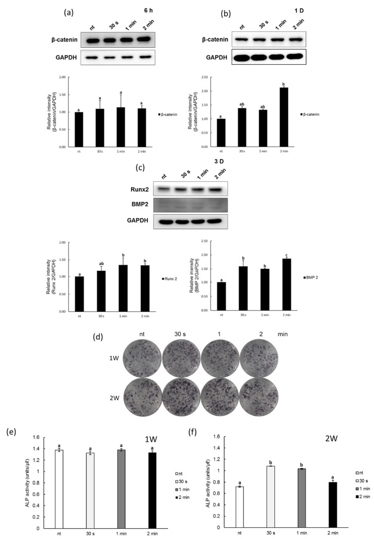
Figure 3.
Effects of NCP on osteogenic differentiation in PDL cells. (a,b) Protein lysates from NCP treated PDL cells were β-catenin, (c) Runx2 and BMP 2 were tested by Western blot assay with specific antibodies at various times. After 1 and 2 weeks, (d) ALP stain and (e,f) activity of PDL cells by NCP treatment were evaluated. Different letters (a, b, c) indicate statistically significant differences by one-way ANOVA (p < 0.05).

