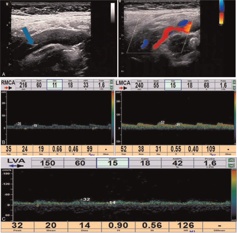
Figure 2.
Cervical vascular ultrasound and transcranial Doppler (TCD) examination. A. Cervical vascular ultrasound showed a membrane structure with moderate echo in the lumen of the right common carotid arteries, pulsating with the blood flow. Low echo could be detected in the lumen of the lesion, resulting in local severe stenosis of the common carotid artery and distal occlusion of the internal carotid artery. The initial diagnosis was carotid arterial dissection with intramural hematoma. B & C. The pulsation of bilateral middle cerebral arteries was low, especially on the right side, and the low velocity blood flow signal was detected in the left vertebral artery, with relatively high resistance changes. LVA = left vertebral artery.

