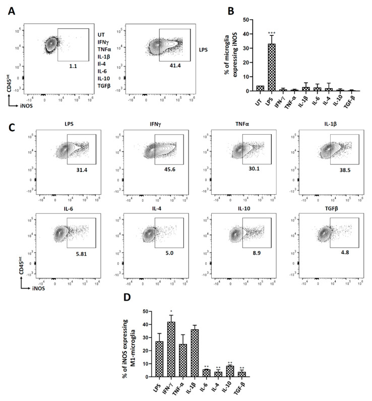
Figure 6.
iNOS expression in microglial cells. Microglial cells were cultured from d7-10 pups as described previously, treated with select pro- and anti-inflammatory cytokines, and analyzed for iNOS expression after 24 h of treatment. Microglial cells polarized to the M1 state were also treated with various pro- and anti-inflammatory cytokines for 24 h followed by assessment of iNOS expression. (A) Representative contour plot of microglial cells (CD45int) expressing iNOS after treatment with various pro- and anti-inflammatory mediators. (B) Representative graph showing the expression of iNOS after treatment with different pro- and anti-inflammatory mediators. (C) Contour plots and (D) graphical representation of iNOS expression on M1-polarized microglia after 24 h of treatment with select pro- and anti-inflammatory cytokines. UT-untreated control. Experiments were carried out three times with duplicate wells. * p < 0.05; ** p < 0.01; *** p < 0.001.

