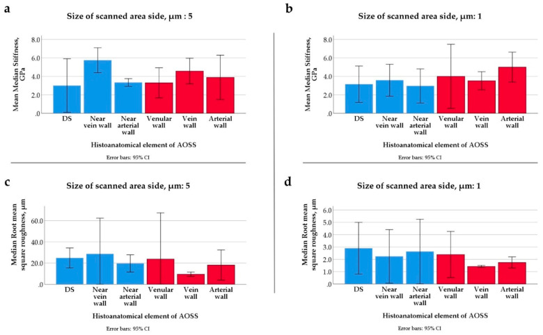
Figure A8.
Physical properties of the histoanatomical elements of dehydrated LS-ECM (in CE liver AOSSs) measured in AFM scans with areas 5 × 5 µm (a,c) and 1 × 1 µm (b,d). The elements of the parenchymal compartment are shown by blue bars, and the elements of the stromal compartment are shown by red bars. (a,b) Median stiffness and (c,d) root mean square roughness. No statistically significant differences of stiffness and roughness were found between the studied histoanatomical elements at both AFM imaging scales (5 × 5 and 1 × 1 µm) in the given set of samples (N = 18 for each scanned area size). Abbreviations: DS—Disse’s space; “Near vein wall” and “Near arterial wall,” the properties of the areas of the parenchymal compartment measured in vicinity to veins and arteries, respectively.

