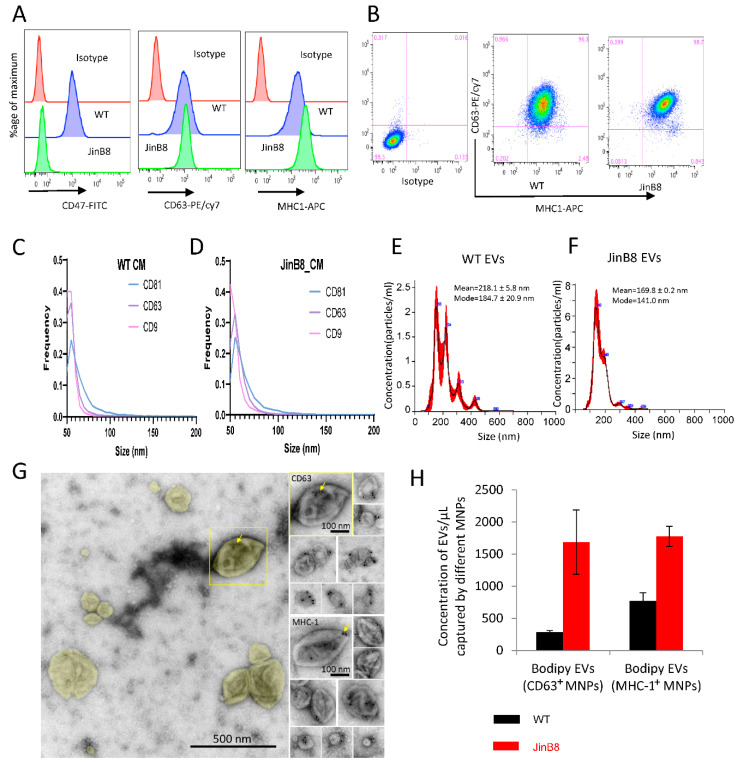
Figure 1.
(A,B) WT Jurkat and JinB8 T cells were stained using conjugated CD47, CD63 and MHC-I antibodies and analyzed by flow cytometry. (C,D) NanoView analysis of size and expression of the tetraspanins CD9, CD63, and CD81 on EVs in concentrated conditioned media derived from WT Jurkat and JinB8 cells. (E,F) Size and concentration of EVs purified from WT and JinB8 cells were measured using Nano Sight. (G) Negative stain EM of immunogold labeled EVs purified from activated Jurkat T cells. Left, overview of EVs colorized yellow in a typical field of view. The boxed EV is immunogold labeled for CD63 and shown to the upper right in a montage with other CD63-labeled EVs. Lower right, a montage of EVs immunogold labeled for MHC-1. Arrows indicate 10-nm immunogold particles. (H) Quantification by flow cytometry of CD63+ and MHC-I+ EVs derived from WT Jurkat and JinB8 cells purified using size exclusion chromatography. Data are representative of two independents biological experiments.

