Figure 5.
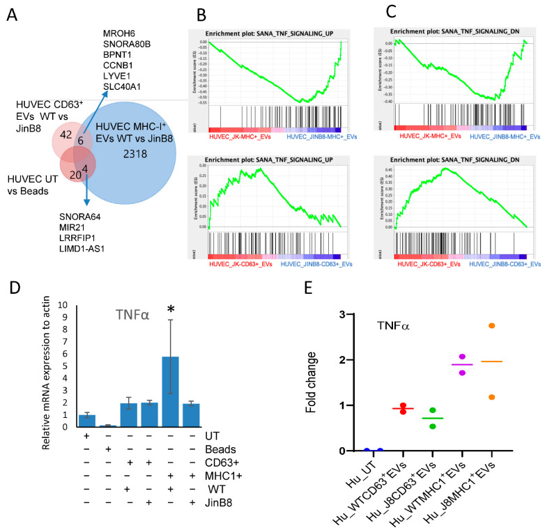
Immunocaptured CD63+ and MHC-I+ EVs as in Figure 3A were co-cultured with HUVEC for 3 days, and differential mRNA expression was evaluated using microarray analysis. (A) Venn diagram showing comparative analysis of CD47-dependent transcripts (p < 0.05) in HUVEC treated with beads alone or treated with CD63+ or MHC-I+ EVs from WT Jurkat or JinB8 T cells. (B,C) Geneset enrichment analysis plots showing enrichment of a TNF signaling signature in HUVEC treated with CD63+ EVs from WT Jurkat vs. JinB8 T cells (B) or MHC-I+ EVs from WT Jurkat vs. JinB8 T cells (C). (D) TNFα mRNA expression was validated via real-time PCR with ß-actin as control (1 biological replicate). (E) The increase in fold change of secreted TNFα in conditioned medium from HUVEC treated with the indicated EVs for 3 days. Secreted TNFα was undetectable from supernatant of untreated HUVEC cells, but control bead-treated sample had background values. Therefore, results are presented as fold change with respect to background value (n = 2). Supplemental Data files related with Figure 5; /Data/s3A, /Data/s3B, /Data/s2C. * p < 0.05.

