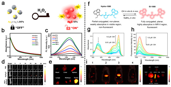
Figure 4.
(a) Schematic representation of the mechanism of H2O2-activatable nanoprobe Ag/Ag2S JNPs for NIR-II fluorescence imaging in vivo. (b) Electronic absorption and (c) fluorescence spectra of Ag/Ag2S JNPs after treatment with H2O2 at different time points. (d) NIR-II fluorescence imaging of tumor-bearing mice at different time points after Ag/Ag2S JNP injection in vivo. (e) NIR-II fluorescence imaging of tumor and main organs excised from the Ag/Ag2S-treated mice group. Reproduced with permission [82]. Copyright 2021, American Chemical Society. (f) Chemical structures and mutual conversion processes of Hydro-1080 and Et-1080. (g) Electronic absorption and (h) fluorescence spectra of Hydro-1080 after reaction with different concentrations of ·OH. (i) NIR-IIa fluorescence imaging of mice injected first with different doses of APAP (0, 300, 500, 500 mg/kg) and then with Hydro-1080 (1 mM, 5 mL/kg). In addition, the fourth mouse in the picture was intraperitoneally pre-injected with ABT (100 mg/kg) 24 h before injecting APAP. 1: Hydro-1080; 2: APAP (300 mg/kg) + Hydro-1080; 3: APAP (500 mg/kg) + Hydro-1080; 4: APAP (500 mg/kg) + Hydro-1080 + ABT (100 mg/kg). (j) NIR-IIa fluorescence imaging of main organs and tissues of mice injected with APAP (500 mg/kg) and Hydro-1080. Reproduced with permission [88]. Copyright 2019, American Chemical Society.

