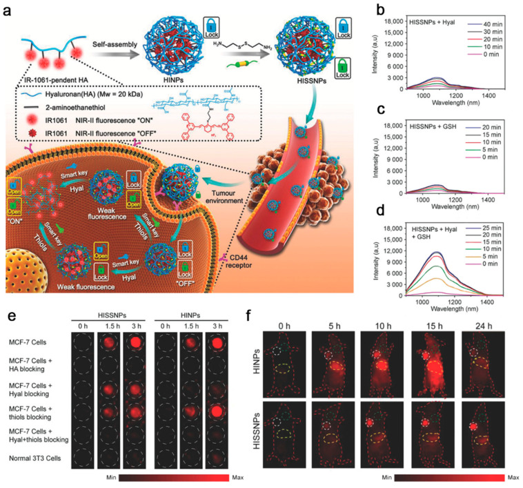
Figure 10.
(a) Schematic diagram of the synthetic route of dual-lock-and-key-controlled NIR-II fluorescence probe HISSNPs and its corresponding mechanism for tumor-specific imaging. Time-dependent fluorescence (excited at 808 nm) spectra of HISSNPs incubated with (b) Hyal, (c) GSH, and (d) Hyal and GSH in PBS. (e) NIR-II fluorescence imaging of 3T3 cells and MCF-7 breast cancer cells at the time points of 0 h, 1.5 h, and 3 h after different treatments. (f) Time-dependent NIR-II fluorescence imaging of MCF-7 cancer xenografts model mice after injection with HISSNPs and HINPs (35 mg/kg). White circle: tumor site; yellow circle: abdominal liver site; green circle: muscle (refers to normal tissue). Reproduced with permission [139]. Copyright 2018, WILEY-VCH.

