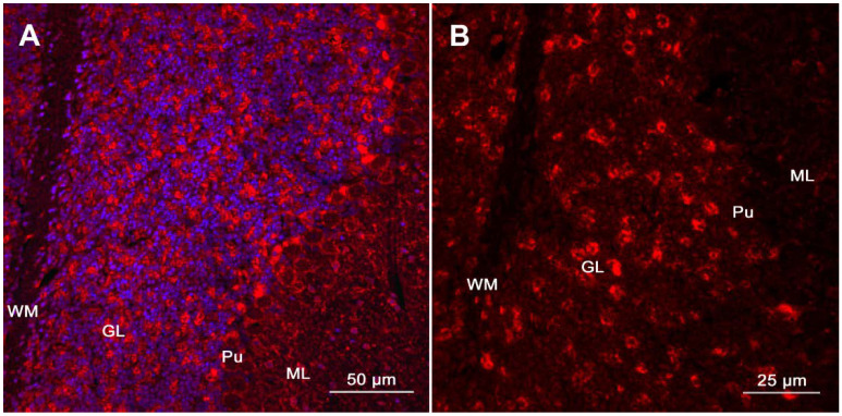
Figure 1.
Detection of specific autoantibodies using tissue-based immunofluorescence. (A). Immunoreactivity of a patient’s CSF with sagittal mouse brain sections is demonstrated via TIF (Cerebellum, magnification: 100×, counterstaining of nuclei by Hoechst 33342 at 1:10,000). (B). Patient’s antibodies show a prominent immunolabeling of the neuropil of the granular layer (GL), whereas binding to the white matter (WM), Purkinje cell layer (Pu), and the molecular layer (ML) is less evident (magnification: 400×). This staining pattern is specific for neurexin-3alpha-antibodies and has been described in the first report of neurexin-3alpha-antibody associated autoimmune encephalitis [108]. Presence of antibodies targeting neurexin-3alpha (a synaptic protein) in patient’s CSF was subsequently confirmed via CBA (Figure adapted from Loehrer et al. [109], reproduced with permission from John Wiley and Sons).

