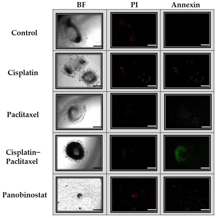
Figure 5.
Visual display of apoptosis and necrosis of Caov-3 spheroids on day 7. Caov-3 spheroids were treated and imaged on day 7 with green annexin V staining (apoptosis) and red propidium iodide (PI) staining (necrosis), as well as under bright field microscopy (BF). Under control conditions, after 7 days, the spheroids showed little necrosis and no apoptosis. Spheroids exposed to single-agent chemotherapy (cisplatin alone, paclitaxel alone) demonstrated the same low level of apoptosis and necrosis as the control. Those spheroids exposed to cisplatin–paclitaxel demonstrated the brightest apoptosis staining and little necrosis staining. Spheroids treated with panobinostat had little cellular material left due to the cytotoxicity of the drug; however, what is present demonstrates high levels of apoptosis and necrosis. Scale bar is 100 µm (n = 2).

