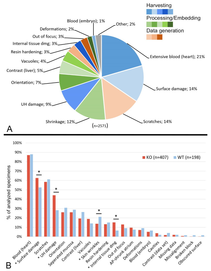
Figure 1.
Frequency of artefacts. (A) Pie chart of artefact types relative to the total number of described artefacts. Harvesting artefacts in blue, processing/embedding artefacts in green, data generation artefacts in orange. “Shrinkage” includes extensive skin wrinkles, shrunk cardiac atrium and separated stomach mucosa; “UH damage” includes damaged and removed umbilical hernia and damaged intestinal slings; “Internal tissue damage” includes tissue damage with or without surface damage. (B) Bar graph of artefact types relative to total number of analyzed specimens, comparing wild type and mutants. Asterisk indicates artefact type with significant difference between KO and WT embryos (* p < 0.05). Abbreviations: AP: anterior-posterior; KO: knockout; WT: wildtype; UH: umbilical hernia.

