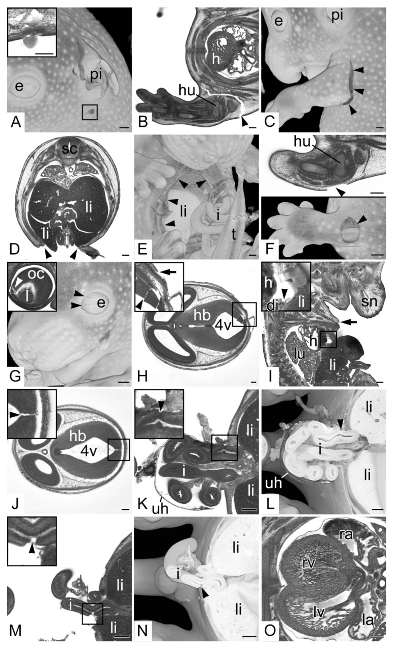
Figure 2.
Specimen harvesting artefacts. (A) Semitransparent volume model of the head from the left. Pinna (pi) broken. Box indicating puncture damage. Inlay: transverse HREM section showing puncture damage. (B,C) Torn upper left extremity (arrow heads), (B) transverse HREM section, ventral to the left (C) 3D model from the left. (D,E) Ruptured ventral body wall (arrow heads) liver (li) protruding from abdominal cavity, (D) transverse HREM section, ventral on bottom, (E) semitransparent volume model from ventral. Note the broken tip of the tail (t). (F) Circumscribed damage of the skin of the left arm (arrow head), transverse HREM section on top, semitransparent volume model on bottom. Note the underlying muscle visible in the 3D model. (G) Damaged eyeball (e) (black arrow heads), semitransparent volume model from the left. Inlay: 2D section showing teared optic cup (oc) (white arrow head). (H) Damage of hindbrain (hb) (arrow head) in combination with surface damage (arrow), transverse HREM section ventral to the left. (I) Damage of the diaphragm (di) combined with ruptured body wall (arrow), sagittal resection ventral to the right. (J) Hindbrain (hb) defect (arrow head) in embryo with intact surface, transverse HREM section, ventral to the left. (K,L) Torn intestine (i) (arrow head) in embryo with intact wall of umbilical hernia (uh). (K) Transverse HREM section, ventral to the left. (L) Semitransparent volume model transected at height of (K). (M,N) Torn intestine (i) (arrow head) in embryo with damaged wall of umbilical hernia. (M) Transverse HREM section, ventral to the left. (N) Semitransparent volume model transected at height of (M). (O) Heart partly filled with blood. Note the difference in visibility of the internal structures of the blood-filled right ventricle (rv) and right atrium (ra) compared to the empty left ventricle and atrium (lv,la). Abbreviations: 4v: 4th ventricle, di: diaphragm, e: eye, h: heart, hb: hindbrain, hu: humerus, i: intestine, l: lens, la: left atrium, li: liver, lv: left ventricle, oc: optic cup, pi: pinna, ra: right atrium, rv: right ventricle, sc: spinal cord, sn: snout, t: tail, uh: umbilical hernia. When not stated otherwise, box indicates magnification in inlay. Scale bars = 250 µm.

