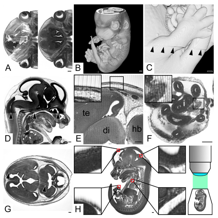
Figure 5.
Data generation artefacts. (A) Original HREM section image partly obscured by unremoved section. (B) Volume model of embryo missing images from top of the head (C) Misalignment between stacks of consecutive HREM images (arrow heads). A 3D model from right. (D) Change of image contrast (arrow heads) at the level of the hard palate. Sagittal resection, ventral to the right. (E,F) Straight (E) and wave-like (F) lines perpendicular to the cutting direction in original HREM images. (G) Scratches in original HREM sections running in parallel to the cutting direction. (H) “Bleeding through”. Sagittal resection, ventral to the left. Inlays show magnifications of areas inside the red boxes. The cranial borders of darkly stained structures appear as fading into cavities in the direction of the image capturing optics. Note the scheme of the optical setup indicating that the embryo is imaged from cranial to caudal. Abbreviations: di: diencephalon, hb: hindbrain, i: intestine, te: telencephalon. Scale bars = 250 µm.

