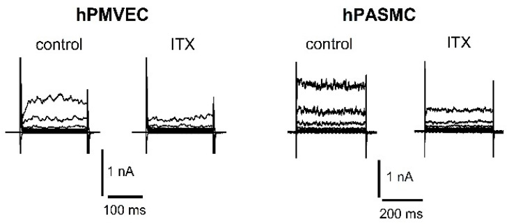
Figure 2.
Representative patch-clamp recordings of K+ currents in primary human pulmonary microvascular endothelial cell (hPMVEC) and primary human pulmonary arterial smooth muscle cell (hPASMC). Recordings show control whole-cell K+ current and the reduction of the current after application of 100 nM ITX. In hPMVEC, currents were evoked from a holding potential of −50 mV using 200-ms voltage steps from −70 up to +90 mV in +20 mV increments. In hPASMC, currents were evoked from a holding potential of −60 mV using 400-ms voltage steps from −80 up to +80 mV in +20 mV increments. Experimental solutions have been described previously [47].

