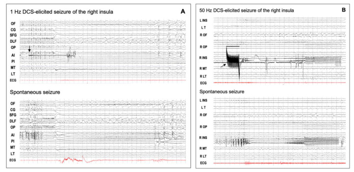
Figure 4.
DCS-induced seizures. (A) Low-frequency (1 Hz) DCS-induced and spontaneous insular seizures. This 18-year-old male patient had seizures characterized by an explosive hypermotor behavior without any aura. From the start of stimulation (arrow), the elicited discharge was quite visible between the DCS artifacts, and its spatio-temporal organization was quite similar to that of the spontaneous seizure. OF: orbito-frontal cortex; CG: cingulate gyrus; SFG: superior frontal gyrus; DLF: dorsolateral frontal cortex; OP: opercular cortex; AI: anterior insula; PI: posterior insula; MT: mesio-temporal structures; LT: lateral temporal cortex; ECG: electrocardiogram. (B) High-frequency (50 Hz) DCS-induced and spontaneous insular seizures for the same patient as in Figure 2. The right postero-superior insular DCS (arrow) induced a seizure which showed the same time evolution and spatial organization as that of the spontaneous discharge, except that the initial part of the elicited discharge was masked by DCS artifacts. DCS: direct cortical stimulation; L: left; R: right: INS: insula; T: temporal lobe; OF: orbito-frontal cortex; OP: opercular region; MT: mesio-temporal structures; LT: lateral temporal cortex; ECG: electrocardiogram. These EEG recordings can be viewed in a larger size in the supplemental material (Figure S1).

