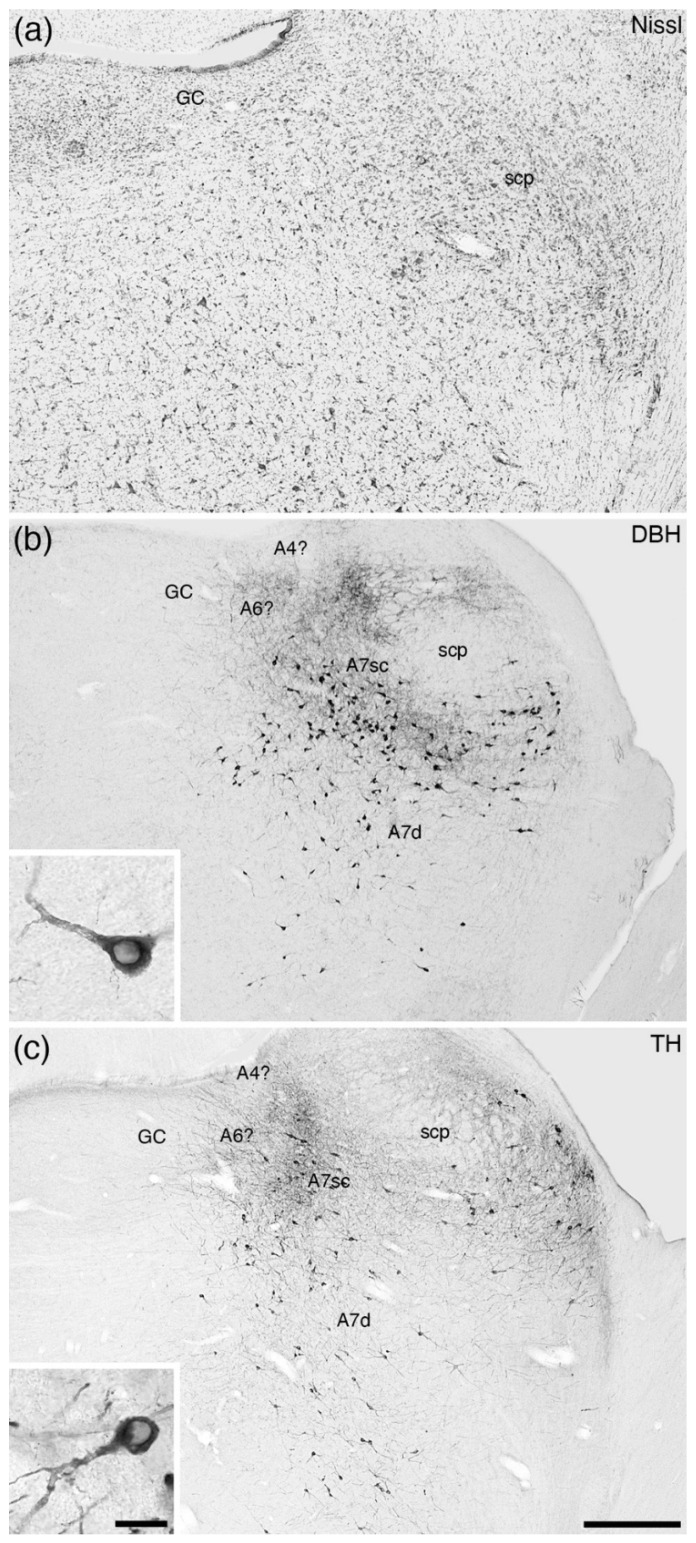
Figure 4.
Low-magnification photomicrographs of the subcoeruleus of the tree pangolin [25], revealed with dopamine-β-hydroxylase (DBH, (b)) and tyrosine hydroxylase (TH, (c)) immunostaining, to compare with an adjacent Nissl-stained section (a). Note the presence of a larger-than-usual compact portion of the subcoeruleus (A7sc) and the diffuse portion of the subcoeruleus (A7d). No apparent dorsolateral division of the locus coeruleus (A4?) or locus coeruleus proper (A6?) is observed within the periventricular grey matter of the rostral hindbrain (GC). The tree pangolin is the only species in which the absence of a locus coeruleus, A6, has been observed [25]. Insets in (b,c) show a high-magnification image of the neurons that form the A7d. In all images, dorsal is to the top and medial to the left. Scale bar in (c) = 500 µm and applies to all images. Scale bar in inset c = 25 µm and applies to both insets. scp—superior cerebellar peduncle.

