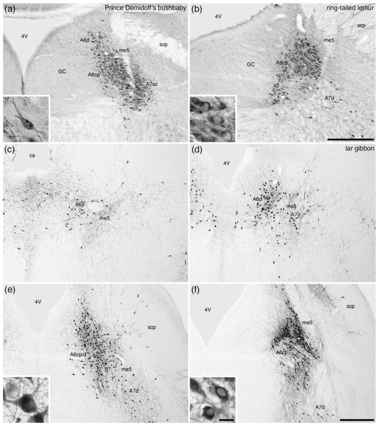
Figure 7.
Low-magnification photomicrographs of the locus coeruleus (A6), revealed with tyrosine hydroxylase immunostaining, in the brains of three species of primates, (a) Prince Demidoff’s bushbaby (Galago demidoff) [52], (b) ring-tailed lemur (Lemur catta) [52], and a rostro-caudal series, with each image being approximately 1 mm apart, through the A6 of the lar gibbon (Hylobates lar) [58]. Note the presence of both diffuse (A6d) and compact (A6cp, compact portion of the locus coeruleus, primate-type) portions of the A6 in primates, with the caudal end of the A6 (d) showing the region of highest density of immunostained neurons. Insets in (a,b,e,f) show a high-magnification image of the neurons from the A6d (a,e) and A6cp (b,d). In all images, dorsal is to the top and medial to the left. Scale bar in (b) = 500 µm and applies to (a,b). Scale bar in (f) = 1 mm and applies to (c–f). Scale bar in inset (f) = 25 µm and applies to all insets. 4V—fourth ventricle; ca—cerebral aqueduct; GC—periventricular grey matter of the rostral hindbrain; me5—fifth mesencephalic tract; scp—superior cerebellar peduncle.

