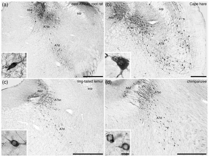
Figure 11.
Low-magnification photomicrographs of the subcoeruleus, or A7, region revealed with immunostaining for tyrosine hydroxylase in four mammalian species belonging to the Euarchontoglires superorder of Eutherian mammals, including (a) the east African root-rat (Tachyoryctes splendens) [pers. obs.], (b) Cape hare (Lepus capensis) [50], (c) the ring-tailed lemur (Lemur catta) [52], and (d) the chimpanzee (Pan troglodytes) [58]. Note the presence of the compact portion of the subcoeruleus (A7sc) in the dorsal aspect of the parvicellular reticular nucleus, with scattered more widely distributed neurons throughout the parvicellular reticular nucleus forming the diffuse portion of the subcoeruleus (A7d). Insets in each image show a high-magnification image of the neurons that form the A7d in each species. In all images, dorsal is to the top and medial to the left. Scale bars in (a,b) = 500 µm and apply to the respective images. Scale bars in (c,d) = 1 mm and apply to the respective images. Scale bar in inset (d) = 25 µm and applies to all insets. scp—superior cerebellar peduncle.

