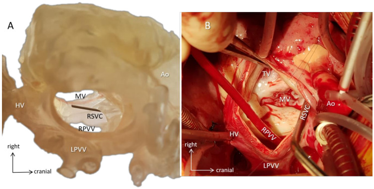
Figure 8.
Surgical simulation. Case 13 with dextrocardia (mirror-image arrangement, bilateral SVCs, hemiazygos continuity of interrupted IVC, common atrium, incomplete AV defect, valvar pulmonary stenosis, vascular ring). (A) A 3D-printed hollow model viewed from the orientation of the surgeon standing on the left side of the patient demonstrates the intracardiac anatomy. A probe emerges in the mouth of the right superior vena cava (RSVC). By identifying anatomical landmarks, e.g., the AV valves and the entrances of the pulmonary and hepatic veins, surgical steps can be simulated, and size and shape of the baffle can be designed preoperatively. (B) Intraoperative representation of the same anatomy. The surgeon identifies structures already familiar with from the 3D model (e.g., metal suction tube is in the right superior vena cava), and the course of the operation progresses along with the preoperative plans. The 3D model and the intraoperative image are closely matched. Abbreviations: Ao: aorta, HV: hepatic veins, LPVV: left-sided pulmonary veins, MV: mitral valve, RPVV: right-sided pulmonary veins, RSVC: right superior vena cava, TV: tricuspid valve.

