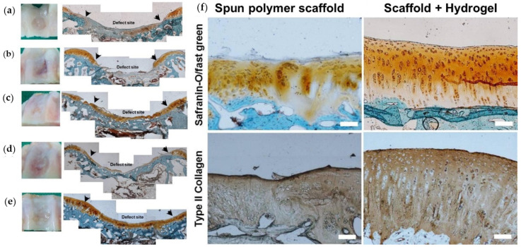
Figure 6.
Photographs of the defect site and histological staining (safranin-O/fast green) of cryo-sectioned samples in rabbit knee at 4 months. (a) Control partial defect site; (b) electrospun polymer scaffold with no cell impregnation; (c) the same scaffold with cells; (d) the polymer scaffold/hydrogel without cells; and (e) the scaffold–hydrogel composite with cells. The arrows point to the borders of initial defect site. Finally, (f) photographs of histological (safranin-O/fast green) and immune-histochemical (type II collagen) stained images of cryo-sectioned samples in rabbit knee at 4 month after implantation of electrospun scaffolds or scaffold–hydrogel composites are shown with scale bars: 100 μm. Reproduced from [114] with permission.

