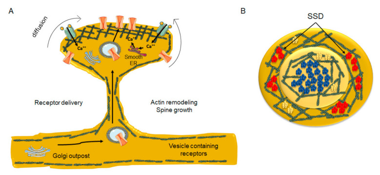
Figure 4.
Diagrammatic depiction of dendritic spine remodeling during synaptic plasticity and associated neurotransmitter receptor clustering in nanodomains. (A) remodeling of the submembrane actin meshwork and incorporation of newly synthesized/laterally diffusing receptors from non-active areas of the spine head and neck into the PSD area, thereby increasing neurotransmitter receptor number at the site of contact with presynaptic boutons. At the crest of the spine receptors become entrapped by actin corrals, which might also present a lipid composition distinct from the bulk lipid bilayer. Golgi outpost in the dendrite and satellite Golgi outposts are found in the spine head, as well as smooth ER outposts. (B) Top view of the PSD. NMDARs (blue) are predominantly located at the center of the PSD in a single nanocluster, whereas AMPARs (red) are segregated into several nanodomains (sub-synaptic domains, SSD) surrounding the central NMDAR nanodomain. In contrast, mGluR5 (yellow) are aggregated into small clusters or homogeneously distributed at the PSD.

