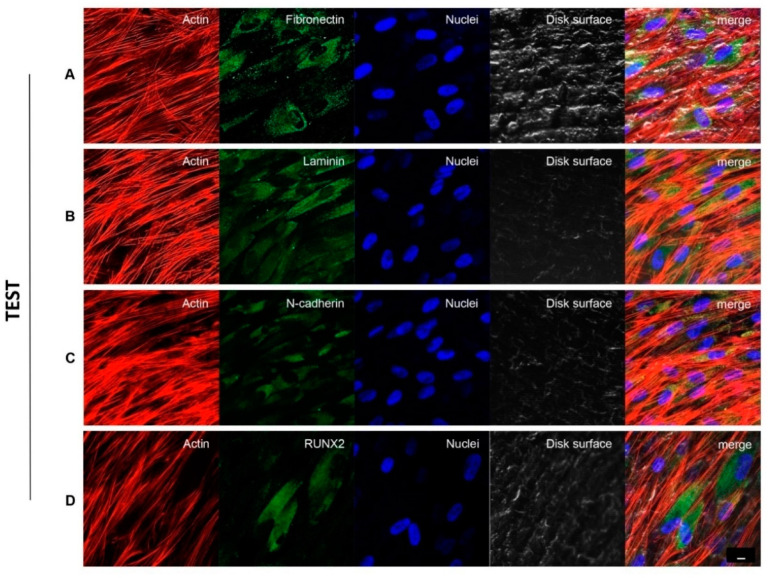
Figure 4.
hPDLSCs cultured on TEST titanium implant surface observed after 8 weeks. Cytoskeleton actin was stained in red fluorescence; specific markers: Fibronectin (A), Laminin (B), N-cadherin (C) and RUNX2 (D) were stained in green fluorescence; nuclei were stained in blue fluorescence. Scale bar: 10 µm.

