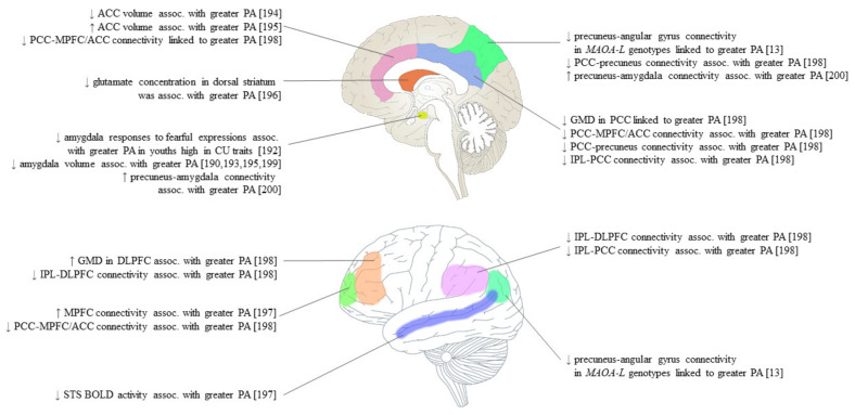
Figure 2.
Neuroimaging findings of proactive aggression (PA), as quantified by the Reactive–Proactive Questionnaire [11], grouped by brain region. ACC, anterior cingulate cortex; Assoc., associated; PCC, posterior cingulate cortex; MPFC, medial prefrontal cortex; CU, callous–unemotional; MAOA-L, monoamine oxidase A low-activity genotype; GMD, grey matter density; IPL, inferior parietal lobe; DLPFC, dorsolateral prefrontal cortex; STS, superior temporal sulcus; BOLD, blood oxygenation level-dependent.

