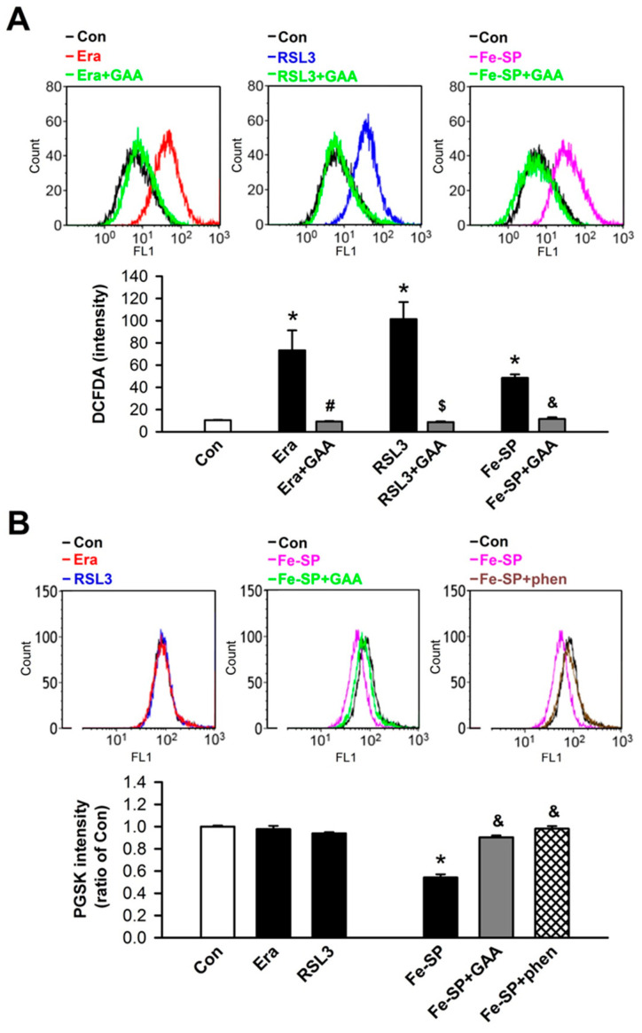
Figure 3.
GAA reduces ROS and iron accumulation in H9c2 cardiomyoblast cells. (A) Treatment with 1-μM Era, 0.1-μM RSL3, and 0.5-μM Fe-SP for 10 h significantly enhanced intracellular ROS in H9c2 cells; these effects were decreased by GAA (2 μM). Top panel: intracellular ROS levels were assessed by flow cytometry analysis using a CM-H2DCFDA probe (an indicator of intracellular ROS); bottom panel: quantitative data (n = 4). (B) Fe-SP, but not Era or RSL3, significantly increased intracellular iron content; this effect was abolished by 2-μM GAA or 50-μM 1, 10-phenanthroline (phen; a ferrous iron chelator). The bottom panel shows data obtained from analysis of the top panel (n = 4). For each flow cytometric analysis, 10,000 cells were counted and plotted. Data are shown as overlapping histogram plots. The same experimental group was assessed by repeating cell culture 4 times. All data represent means ± SEM. * p < 0.05 versus the control group; # p < 0.05 versus Era; $ p < 0.05 versus RSL3; & p < 0.05 versus Fe-SP. Con, control; Era, erastin; Fer-1, ferrostatin-1; Lip-1, liproxstatin-1; RSL3, (1S,3R)-RSL3; Fe-SP, chlorido [N, N-disalicylidene-1,2-phenylenediamine] iron (III); GAA, gossypol acetic acid; phen, 1, 10-phenanthroline.

