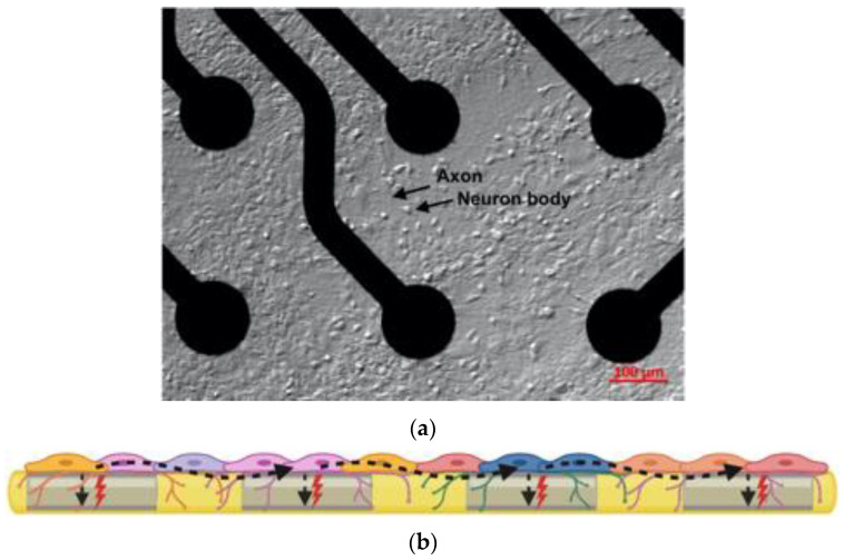
Figure 1.
(a) hCNS-Neuron Cells culture on an MEA well, on the top of the recording electrodes; the black arrows highlights an axon and a cellular body, 10 magnification; (b) a schematic “cross-sectional” overview of the MEA plate technology: spontaneous extracellular action potentials or spikes (red thunders) originated by different neuron types are recorded by the electrodes (gray rectangles). Besides the spontaneous cellular spikes registered by a single or multiple electrodes, the electrochemical signal can also travel across the neural network, indicating synaptic communication (denoted by undulated black dashed arrows).

