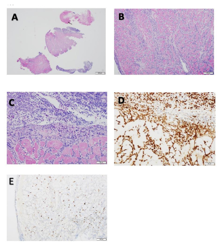
Figure 3.
(A) Hematoxylin and eosin stain (low-power view, scale: 500 µM ) showing dense lymphocytic infiltrates involving every piece of the tissue biopsy sample and endocardium. (B) Hematoxylin and eosin stain of the dense lymphocytic infiltrates (midpower view, scale: 100 µM). The infiltration is widespread around each myocyte. (C) Hematoxylin and eosin stain of the infiltrating cells (high-power view, scale: 50 µM) showing highly malignant features, including numerous apoptotic bodies, frequent mitoses, and myocyte necrosis. (D) Immunohistochemical stain for PAX-5 demonstrating diffuse and strong positivity for the marker (higher-power view, scale: 50 µM). (E) Immunohistochemical stain for TdT, a marker for diagnosis of acute leukemia. About 10% of the tumor cells were positive for TdT, supporting the diagnosis ALL. No features of sarcoidosis or amyloidosis are shown (midpower view, scale: 100 µM).

