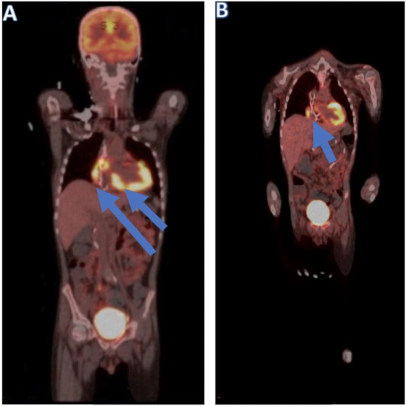
Figure 4.
PET scans of the patient showing 18F-fluorodeoxyglucose uptake in the myocardium involving the right and left ventricles. (A) The arrows indicate abnormal uptake in the myocardium involving both ventricles, with a maximum standard uptake value of 15. This uptake is not a typical finding as leukemia in consideration of the high metabolic activity of the lesions. (B) The arrows indicate persistent hypermetabolism involving the myocardium predominantly in the lateral wall of the right atrium, the right ventricle, and into the ventricle septum, with a maximum standard uptake value of 8.7.

