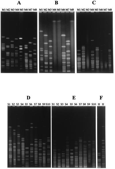
FIG. 1.
Analysis of PFGE patterns of M. catarrhalis, S. pneumoniae, and H. influenzae. M. catarrhalis isolates were classified as M1, M2, M3, M4, M5, M6, M7, or M8 based on their PFGE patterns with SpeI (A), NotI (B), and NheI (C). S. pneumoniae isolates were classified as S1, S2, S3, S4, S5, S6, S7, S8, S9, or S10 based on their PFGE patterns with SmaI (D) and ApaI (E). All H. influenzae isolates had the same PFGE pattern with SpeI (F, left lane) and SmaI (F, right lane).

