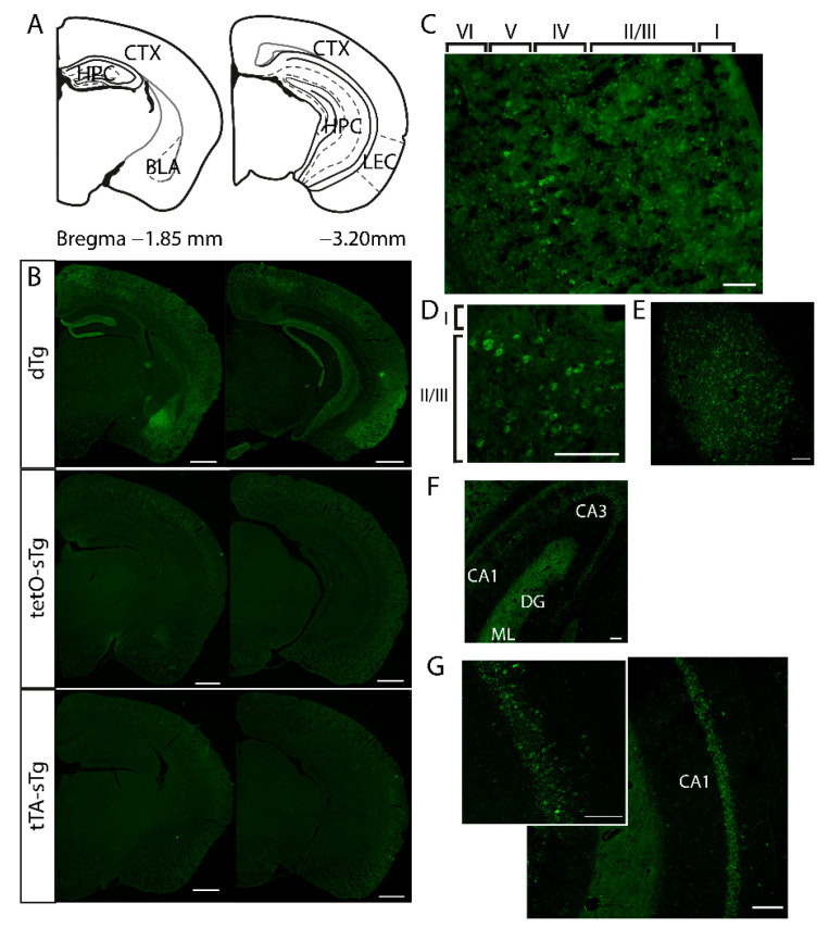
Figure 1.
The basal expression of mPAK3-GFP proteins in sTg and dTg animals. (A) Schematic of the regions of interest in dorsal and posterior coronal sections. (B) The global expression of mPAK3-GFP proteins in dorsal and posterior coronal half brain sections from dTg and control, tetO-sTg, and tTA-sTg mice. Scale bar = 1000 μm. (C–G) Higher magnifications of the regions of interest in dTg mice. Expression of mPAK3-GFP proteins can be seen in (C) all cortical layers, including the (D) LEC, as well as the (E) BLA, and (F) hippocampal CA3 and (G) CA1 regions. Scale bars: 100 μm. CTX, cortex; HPC, hippocampus; BLA, basolateral amygdala; LEC, lateral entorhinal cortex; dTg, double transgenic; tetO-sTg, tetO-mPAK3-GFP single transgenic; tTA-sTg, Fos-tTA single transgenic; CA1, cornus ammonis 1; CA3, cornus ammonis 3; DG, dentate gyrus; ML, molecular layer; GCL, granule cell layer.

