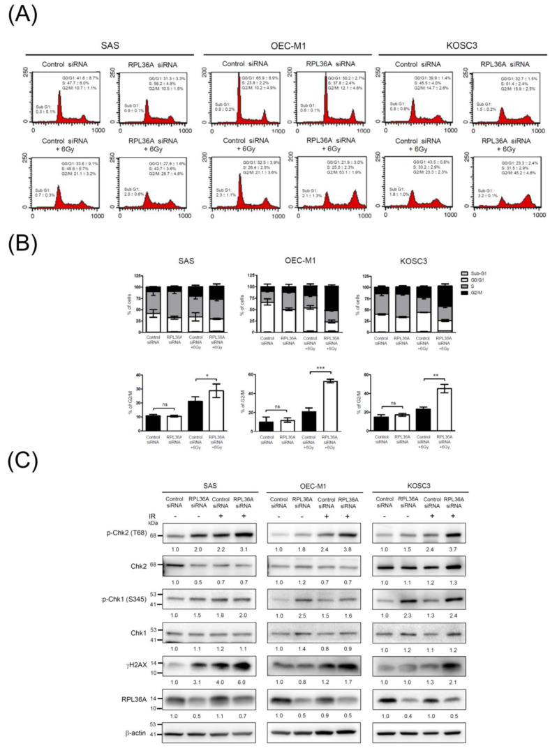
Figure 4.
RPL36A depletion sensitizes cells to DNA damage and promotes G2/M cell cycle arrest in response to irradiation: (A) SAS, OEC-M1, and KOSC3 cells were transfected with control siRNA and RPL36A-specific siRNA. Cells were simultaneously subjected to 6 Gy irradiation, flow cytometry analysis, and Western blotting. After irradiation for 48 h, the DNA content was determined by PI staining followed by flow cytometry analysis; (B) the bar charts show the distribution of the transfected cells in the Sub-G1, G1, S, and G2/M cell cycle phases. Quantification analysis of the cell cycle with G2/M distributions acquired from three independent experiments. Mean values of three independent experiments ± SD are shown. The p-values were calculated by using unpaired Student’s t-test. ns, nonsignificant, * p < 0.05, ** p < 0.01, and *** p < 0.001; (C) total cellular proteins were subjected to Western blotting and analyzed using the indicated antibodies. β-Actin was used as an internal control. The values represent the quantified signals of Western blotting obtained from indicated antibodies and normalized to those of β-actin.

