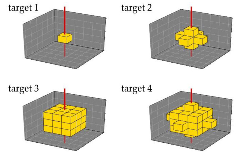
Figure 2.
All four different targets to simulate XFI-scans in the thorax. Shapes shall represent a tumor or inflammation at different sizes and consist of voxels of lung-tissue, to which certain amounts of gold were added. In reality, such an area would have a round shape; targets follow voxel geometry of voxel phantom, and are thus cubic, depending on their size. Incident beam (red) hits them orthogonally, thus between target 2 and target 3, amount of target voxels in beam volume does not change.

