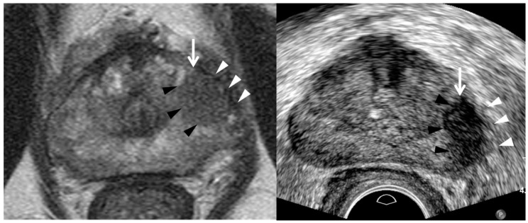
Figure 7.
A 73-year-old man with high PSA (10.71 ng/mL): T2-weighted MR image (left figure) shows a PI-RADS 4 peripheral lesion (white arrow) in the left mid-gland. It is a 1.3 cm hypointense tumor in which the capsule (white arrowheads) is not disrupted, consistent with PI-RADS 4. Transrectal ultrasound image (right figure) shows it is a hypoechoic tumor (white arrow) with irregular or spiculate margin (black arrowheads). The capsule (white arrowheads) neighboring the tumor also appears thick and infiltrative, suggesting extra-capsular extension. Four target cores alone were obtained and systematic biopsy was skipped. The tumor was histologically confirmed adenocarcinoma GS 7 (4 + 3). The tumor shape looks more round on MRI than TRUS. Left peri-prostatic nerves were not saved because extensive capsular extension was detected during the robot-assisted radical prostatectomy.

